

ICCBH2013 Oral Communications Chronic diseases (6 abstracts)
Wnt/β-catenin: a candidate pathway for bone repair in neurofibromatosis type-1
Saber Ghadakzadeh 1, , Saeid Amini Nik 2 , Gurpreet Baht 2 , Heather Whetstone 2 & Benjamin Alman 2,
1Institute of Medical Science, University of Toronto, Toronto, Ontario, Canada; 2Hospital for Sick Children, Program for Developmental and Stem Cell Biology, Toronto, Ontario, Canada; 3Division of Orthopaedics, Department of Surgery, Toronto, Ontario, Canada. *Winner of New Investigator Award
Objectives: Skeletal abnormalities occur in half of the individuals with neurofibromatosis type-1 (NF1), usually in the first 2 years of life; the most difficult to manage is congenital pseudarthrosis of tibia (CPT, tibial non-union) which may lead to the amputation of the limb. Previous work in our lab identified that high levels of β-catenin early in fracture repair, result in a phenotype similar to CPT. There is data showing higher than normal β-catenin levels in CPT samples from NF1 patients, therefore we hypothesized that Wnt/β-catenin pathway mediates the effect of NF1 mutation on bone by altering the differentiation of mesenchymal stromal cells (MSCs) into osteoblasts.
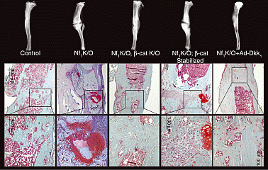
Methods: Nf1fl/fl mice were crossed with conditional β-catenin over-expressing and knock-out models. Adenovirus containing Cre was injected at the site of tibial fractures to both knock-out (k/o) Nf1 and modulate β-catenin levels. Fracture healing was evaluated using radiological, histological and biomechanichal tests.
Results: Nf1−/− tibial fractures were associated with higher cellular β-catenin levels early after fracture and did not heal after 21 days, exhibiting a fibro-cartilaginous phenotype at the site of fracture. Nf1−/−; β-catenin k/o fractures showed repair and union with significantly decreased fibro-cartilaginous tissue. Nf1−/−; β-catenin stabilized fractures did not heal and the amount of fibro-cartilaginous tissue increased significantly. Utilizing Dkk-1, as a specific Wnt/β-catenin signaling inhibitor, at the site of Nf1−/− fractures resulted in markedly improved bone formation and union of fractures.
Consistent with our in-vivo mouse models, human NF1 CPT samples contained increased β-catenin levels. Unlike wild-type bone marrow (BM) MSCs, cultured human NF1 and mouse Nf1−/− BMMSCs showed impaired osteoblastic differentiation which was either rescued or further inhibited by β-catenin signaling down- or up-regulation, respectively.
Conclusion: These data indicate that the level of β-catenin needs to be precisely controlled for normal osteoblastogenesis during fracture repair, and that in NF1 patients, this level is abnormally elevated, inhibiting osteoblastogenesis and bone formation. This study provides important insights into the pathways regulating impaired fracture healing in relation to NF1 patients and will assist the clinical management of non-unions healing.
Volume 2
Article tools
My recent searches
My recently viewed abstracts

Bone Abstracts
ISSN 2052-1219 (online)
© Bioscientifica 2024 |
Privacy policy |
Cookie settings
BiosciAbstracts

Bioscientifica Abstracts is the gateway to a series of products that provide a permanent, citable record of abstracts for biomedical and life science conferences.
Find out more