ICCBH2015 Poster Presentations (1) (201 abstracts)
Association of dental and skeletal fluorosis with calcium intake and vitamin D concentrations in adolescents from a region endemic for fluorosis
Neha Kajale 1 , Prerna Patel 2 , Pinal Patel 2 , Ashish Patel 2 , Bhrugu Yagnik 3 , Rubina Mandlik 1 , Vivek Patwardhan 1 , Vaman Khadilkar 1 , Shashi Chiplonkar 1 , Zulf Mughal 5 , Priscilla Joshi 4 , Supriya Phanse-Gupte 1 & Anuradha Khadilkar 1
1Growth and Endocrine Unit, Hirabai Cowasji Jehangir Medical Research Institute, Jehangir Hospital, Pune, India; 2Department of Biotechnology, Hemchandrachyarya North Gujarat University, Patan, India; 3Maharaja Sayajirao University, Vadodara, India; 4Department of Radiodiagnosis and Imaging, bharati vidyapeeth, Pune, India; 5Department of Paediatric Endocrinology, Royal Manchester Children’s Hospital, Manchester, UK.
Objective: Patan, is a semi urban area in Gujarat, India where fluorosis is endemic (Fluoride concentration in ground water 1.96–10.85 ppm, Patel et al., 2008). Exposure to fluoride is likely to be higher in lower socio-economic class (SEC) due to lack of access to bottled water. Calcium intake and vitamin D status may modify fluorine absorption. Therefore, the aims of this study were to examine: i) prevalence of dental and skeletal fluorosis in children from upper, middle and lower socioeconomic class; ii) association of fluorosis with calcium intake and vitamin D status.
Methods: Cross-sectional study was conducted in 10–14 year apparently healthy adolescents (n=90; boys, n=45), belonging to upper, middle and lower SEC (n=30/SEC, Kuppuswamy scale, 2012) from Patan (Gujrat). Dental fluorosis was graded as mild, moderate, severe by a dentist. Radiographs of right hand and wrist were examined and graded (Teotia et al., 1998). 25OHD and PTH (both using chemiluminescent immunoassay) were measured. Diet was recorded by 24 h recall and calcium intake computed (C-diet 2.1).
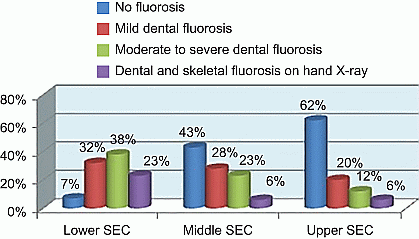
Results: Fluorosis was predominantly seen in lower SEC, (Default 1); there were no skeletal deformities. Mean 25OHD concentrations and dietary calcium were 26.3±4.9, 23.4±4.7, 18.6±4 ng/ml and 406±222, 499±192, 690±245 mg/d respectively for lower, middle and upper SEC (P<0.05). PTH did not vary (P>0.1).79% from upper, 50% from middle and none from low SEC had access to bottled water with appropriate fluoride concentrations(http://www.cdc.gov/fluoridation/faqs/bottled_water.htm). There was significant association between fluorosis and SEC (exponential β=2.5, P<0.01); 25OHD and calcium showed no significant association.
Conclusion: Fluorosis was more common in lower SEC. Relatively adequate calcium intake and 25OHD may have prevented severe skeletal flurosis.
Disclosure: The authors declared no competing interests.

Figure 1 Prevalence (%) of fluorosis in different SEC.


