ICCBH2013 Late Breaking Abstracts (1) (2 abstracts)
Influence of age and gender on spine bone density and TBS microarchitectural texture parameters in infants
Renaud Winzenrieth 1 , Catherine Cormier 2 , Silvana DiGregorio 3 & Luis Del Rio 3
1R&D Department, Med-Imaps, Bordeaux, France; 2Service de Rheumatology A, Hospital Cochin, APHP, Paris, France; 3Cetir Grup Mèdic, Barcelona, Spain.
Children bone knowledge is relatively sparse. This is especially true for infant and for bone microarchitecture data. We have investigated, in this study, the age-related modifications of spine microarchitectural texture, as assessed by TBS, on male and female infants during their two first years of life.
The study group was composed of 143 and 109 healthy female and male infants aged between 0 and 2 years. Height and weight Z-scores were significantly lower than zero (−0.61 and −0.65 for female; −0.62 and −0.77 for male), showing that infants were shorter and underweight relative to The WHO Child Growth Standards. The areal BMD (aBMD) was assessed at spine L1–L4 using a prodigy densitometer (GE-LUNAR, USA). TBS was evaluated using TBS iNsight v2.0 (Medimaps, France). The LMS statistical method proposed by Cole & Green (Stat Med 1992) was used to construct aBMD and TBS age-related curves for each gender using R software (v2.15.3).
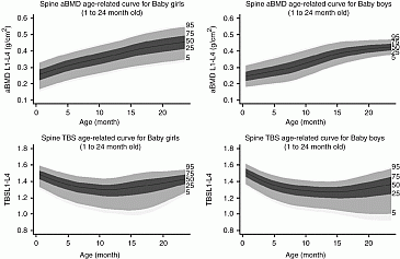
Female and male infant shave the same mean age, height and weight Z-score and TBS (P>0.3) whereas female infants have higher aBMD than male (P=0.01). Before and after 12 months of age, TBS and aBMD correlations in both female and male infants were low (r2<0.20).We have observed a first TBS decrease from birth to 12–15 months (for female and male infants respectively) followed by a TBS increasing without reaching the mean birth TBS value. In parallel, the aBMD always increases (see Fig. 1).

Figure 1 Age-related curves for aBMD and TBS at spine L1-L4 (The black line represents the 50th centile. The dark gray represents the 25th to 75th centiles; The medium gray area represents the 5th 95th centiles; The light gray area represents the 3th to 97th centiles)
To date, our study is the first which has evaluated the modification of spine microarchitectural texture occurring in infants. Results obtained showed similar aBMD and TBS evolution shapes for both male and female infants. Surprisingly, TBS evolution exhibits a decreasing/increasing pattern. We can hypothesis that this pattern correspond to the infants bed-rest phase (no weight loads on the spine→trabecular structure adaptation using minimum energy adaptation rule→TBS decreasing) and the stand/walking phase (weight loads on spine→mechanical stress increasing→trabecular structure positive adaptation→TBS increasing). Further studies have to be conduct to confirm these first findings.
Declaration of interest: R Winzenrieth is a senior scientist at Med-Imaps.
 }
}


