

ICCBH2015 Poster Presentations (1) (201 abstracts)
Dysosteosclerosis from a unique mutation in SLC29A3
Serap Turan 1 , Steven Mumm 2, , Gary S Gottesman 2 , Saygin Abali 1 , Serpil Baş 1 , Zeynep Atay 1 , William H McAlister 4 , Michael P Whyte 2, & *Dr. Turan and Dr. Mumm contributed equally to this work
1Department of Pediatric Endocrinology, Marmara University, Istanbul, Turkey; 2Center for Metabolic Bone Disease and Molecular Research, Shriners Hospital for Children, St Louis, MO, USA; 3Division of Bone and Mineral Diseases, Washington University School of Medicine, St Louis, MO, USA; 4Mallinckrodt Institute of Radiology, Washington University School of Medicine, St Louis, MO, USA.
Dysosteosclerosis (DSS) is the rare osteopetrosis (OPT) distinguished by metaphyseal osteosclerosis with relative radiolucency of widened diaphyses and platyspondyly. In 2012, mutations in the SLC29A3 gene were discovered to cause DSS.
Here, we report a new case of DSS presenting with severe anemia and having a unique homozygous mutation in SLC29A3.
Our patient was the 3rd child of consanguineous Turkish parents. She presented with severe anemia (Hb 3 mg/dl) at age 4 months, and was transfused with a presumptive diagnosis of OPT. After 1 year-of-age, no hematologic abnormality was detected.
Her first fracture occurred at about 2 years-of-age. She underwent surgery several times for bilateral femoral fractures (seven fractures). BMD-DXA L2–L4 z-score was +6 and +8 at 10 and 17 years-of-ages respectively. Mineral homeostasis was intact.
She had no other systemic problems, with normal mentation and good scores in school.
She presented to us at 17 years-of-age. Height was 145.1 cm (<3%), arm span was 159 cm, and upper-lower segment ratio was 0.93. Despite her past femoral fractures, she had a short upper segment and broad shoulders, but no further obvious dysmorphic features except mild prognathism, short philtrum, prominent nose, and small maxillary lateral incisors.
DSS was suspected from reviewing her radiographs taken in early childhood due to her marked metaphyseal sclerosis. Later radiographs showed more diffuse osteosclerosis with ‘sandwich’ vertebrae and platyspondyly.
Sequencing of all the coding exons of SLC29A3 for the proband revealed a novel homozygous 18-bp duplication in exon-3 (c.303_320dup, p.102_107dupYFESYL) resulting in a tandem duplication of 6-amino acids. Both parents were heterozygous for this mutation.
Conclusion: Radiographic findings in DSS evolve during growth and can be misdiagnosed when there is diffuse osteosclerosis as malignant (‘infantile’) OPT.
Disclosure: The authors declared no competing interests.
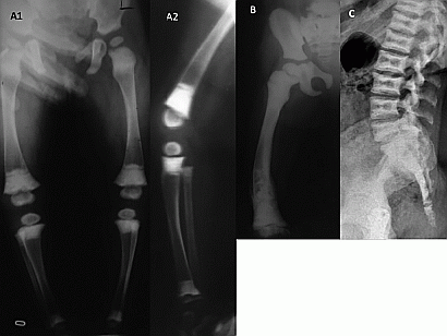
Figure 1 A. Radiographs in early childhood show metaphyseal and epiphyseal scierosis. B. At age 6 years there is diffuse osteoscierosis and flaring in the metaphyses and metadiaphyses, cortical thinning and bowing. C. At age 17 years there is platyspondyly with sandwich vertebrae.
}
Article tools
My recent searches
My recently viewed abstracts
Authors

Bone Abstracts
ISSN 2052-1219 (online)
© Bioscientifica 2026 |
Privacy policy |
Cookie settings
BiosciAbstracts

Bioscientifica Abstracts is the gateway to a series of products that provide a permanent, citable record of abstracts for biomedical and life science conferences.
Find out more